
An atlas of illustrations of pathology (fasc. I-XII)
- Collections
- Anatomie pathologique
- DESCRIPTION
- TABLE DES MATIÈRES
- VOIR PLUS
- Identifiant
- ark:/13685/01866
- Titre
- An atlas of illustrations of pathology (fasc. I-XII)
- Créateur
- New Sydenham Society
- Date
- 1877 – 1899
- Éditeur
- London : The new Sydenham Society
- Siècle
- 19e siècle
- Format
- Nombre de vues : 546
- Source
- Université Paris Cité. BIU Santé Médecine, inv. 1866
- Propriétaire
- Université Paris Cité. BIU Santé Médecine
- Licence
- Licence Ouverte
- Table des matières
-
![0001 - Page sans numérotation - [Plat]](https://numerabilis.u-paris.fr/iiif/2/bibnum:01866:0001/square/200,/0/default.jpg) 0001 - Page sans numérotation - [Plat]
0001 - Page sans numérotation - [Plat]
-
![0002 - Page sans numérotation - [Contreplat]](https://numerabilis.u-paris.fr/iiif/2/bibnum:01866:0002/square/200,/0/default.jpg) 0002 - Page sans numérotation - [Contreplat]
0002 - Page sans numérotation - [Contreplat]
-
![0006 - Page sans numérotation - [Mention d'éditeur]](https://numerabilis.u-paris.fr/iiif/2/bibnum:01866:0006/square/200,/0/default.jpg) 0006 - Page sans numérotation - [Mention d'éditeur]
0006 - Page sans numérotation - [Mention d'éditeur]
-
![0007 - Page sans numérotation - [Page de titre]](https://numerabilis.u-paris.fr/iiif/2/bibnum:01866:0007/square/200,/0/default.jpg) 0007 - Page sans numérotation - [Page de titre]
0007 - Page sans numérotation - [Page de titre]
-
 0009 - Page I - Index
-
 0010 - Page II - Atlas of illustrations of pathology / Contents and index
-
 0011 - Page III - Index
-
 0017 - Page sans numérotation - An atlas of illustrations of pathology / Fasciculus I Diseases of the kidney
-
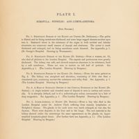 0020 - Page sans numérotation - Plate I / Scrofula : Syphilis : and Lymph-Adenoma. Fig. 1. Scrofulous disease of the kidney and ureter ( Dr. Dickinson). Fig. 2. Scrofulous disease of the kidney (Dr.Sutton) Fig. 3. Scrofulous disease of the kidney (Dr Sutton) . Fig. 4. A mass of syphilitic deposit in the cortical substance of the kidney (Dr Sutton) Fig. 5. Lymph-Adenoma of Kidney (Dr Sutton)
-
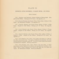 0024 - Page sans numérotation - Plate II / Nephritis after diphtheria; scarlet fever; and burns. Fig.1. Nephritis after diphtheria. Fig. 2. Subacute nephritis after scarlet fever. Fig. 3. Subacute nephritis after scarlet fever. Fig. 4. Acute nephritis after scarlet fever. Fig. 5. Subacute nephritis after scarlet fever. Fig .6. Acute nephritis after a burn. Fig. 7. Acute nephritis after a burn
-
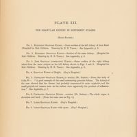 0028 - Page sans numérotation - Plate III / The granular kidney in different stages. Fig.1. Extremely granular kidney. Fig. 2. Extremely granular kidney. Fig. 3. Less granular (contracted) kidney. Fig. 4. Granular kidney of bright. Fig. 5. Contracted granular kidney, in section (Dr. Sutton). Fig. 6. Contracted granular kidney; exterior (Dr. Sutton). Fig. 7. Large granular kidney. Fig. 8. Large granular kidney with cysts
-
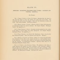 0032 - Page sans numérotation - Plate IV / Embolism : infarction processes from pyaemia : jaundice and purpura : scrofula. Fig.1. Embolic changes in pyaemia (Dr. Sutton). Fig. 2. Embolic changes in pyaemia. Fig. 3. Pyaemic depositsin kidney. Fig. 4. Pyaemic deposits in the kidney. Fig. 5. Results of jaundice and purpura. Fig. 6. A variety of the scrofulous kidney
-
 0035 - Page 1 - Appendix / Plate I
-
 0036 - Page 2 - Appendix / Plate II
-
 0037 - Page 3 - Appendix / Plate II
-
 0038 - Page 4 - Appendix / Plate II
-
 0039 - Page 5 - Appendix / Plate II
-
 0040 - Page 6 - Appendix / Plate II et III
-
 0041 - Page 7 - Appendix / Plate III
-
 0042 - Page 8 - Appendix / Plate IV
-
 0043 - Page sans numérotation - An atlas of illustrations of pathology / Fasciculus II Diseases of the kidney,supra-renal capsules and spleen; with pathological summaries by Dr. Greenfield and Dr. Goodhart
-
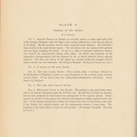 0046 - Page sans numérotation - Plate V / Disease of the kidney. Fig.1. Amyloid disease of kidney in advanced stage. Fig. 2. A section of the same kidney. Fig. 3. The pale flabby kydney. Fig. 4. The same organ seen in section. Fig. 5. Medullary cancer of the kidney
-
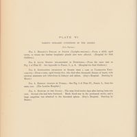 0050 - Page sans numérotation - Plate VI / Various diseased conditions of the spleen. Fig. 1. Hodgkin's disease of Spleen (lympho-sarcoma). Fig. 2. Acute splenic enlargement in diphtheria. Fig. 3. Suppurating infarction of spleen from a case of ulcerative endocarditis. Fig. 4 Embolic changes in pyaemia. Fig. 5. Rupture of the spleen
-
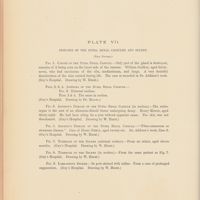 0054 - Page sans numérotation - Plate VII / Diseases of the supra renal capsules and spleen. Fig. 1. Cancer of the supra renal capsule. Fig. 2,3,4 Adenoma of the supra renal capsule. Fig. 3. External surface. Fig. 2 & 4 the same in section. Fig. 5 Addison's disease of the supra renal capsule. Fig. 6. Addison's disease of the supra renal capsule (in section). Fig. 7. Tubercle of the spleen (external surface). Fig. 8. tubercle of the spleen (in section). Fig. 9. Lardaceous spleen
-
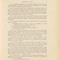 0057 - Page sans numérotation - Plate VIII / Fig. 1. Lardaceous degeneration of the kidney. Fig. 2. Lardaceous degeneration. Fig. 3. Fig. 4 Lardaceous degeneration in earlier stage combined with interstitial fibrous change. Figs. 5. & 6 Lardaceous degeneration (section cornil). Fig. 7. Granular contracted kidney. Fig. 8. From the same
-
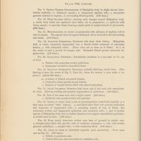 0058 - Page sans numérotation - Plate VIII / Fig. 9. Partial Fibrous degeneration of Malpighian body. Fig. 10 From the same kidney. Fig. 11. Multiplication of nuclet. Fig. 12. Subacute interstitial nephritis. Fig. 13. Scarlatinal nephritis. Fig. 14. Subacute interstitial nephritis. Fig. 15. Acute Catarrhal Nephritis. Fig. 16. Part of the same seen with higher power. Fig. 17. Section of cortex from a case of parenchymatous ( catarrhal) nephritis. Fig. 18. From nearly transverse section near base of pyramid in similar case.Fig. 19. Casts in tubes in interstitial nephritis ( post scarlatinal). Fig. 20. Colloid cast
-
 0061 - Page sans numérotation - Plate IX / Fig. 1. Scarlatinal Nephritis.Fig. 2. Shows two of the glomeruli from the same section as Fig. 1. d', under a higher power. Fig. 3. Section from the same. Part of the wall of a Malpighian body from which the capillary tuft has fallen out. Seen under a higher power. Fig. 4. Scarlatinal Nephritis. Fig. 5. Scarlatinal Nephritis ( From a case fatal 15 months after attack of scarlet fever) Fig. 6. From same kidney as Fig. 5. ,but in a deeper part of cortex, close to medulla . Fig. 7. Subacute interstitial Nephritis. Fig. 8. Chronic Parenchymatous nephritis (large white kidney) with little or no interstitial change
-
 0062 - Page sans numérotation - Plate IX / Fig. 9. Kidney in leucocythaemia. Fig. 10. Swelling of inner cost of small artery in granular contracted kidney.Fig. 11. Tuberculous Pyelo-nephritis. Fig. 12. Fatty degeneration from Alcoholic poisoning. Fig. 13. Fatty degeneration in cancer. Fig. 14. Individual epithelial cells from the preceding section; in various stages of fatty degeneration. Fig. 15. Cystic degeneration of kidney. Fig. 16. From a cyst in kidney near base of pyramid. Fig. 17. Colloid degeneration of kidney. Figs. 18,19,20 and 21 illustrate the hyaline changes found in the splenics arteries in certain febrile conditions. Fig. 18. From a section through the spleen of a case of early scarlatina. Fig. 19. Artery in longitudinal section. Fig. 20. Malpighian corpuscle from the spleen a case of early scarlatina, showing three different zones, a, b, c. Fig. 21. Part of the central and intermediate zone of the same Malpighian corpuscle as in Fig. 20.,only more highly magnified (180 diam.) Fig. 22 Hodgkin's disease. Fig. 23. Adenoma of the supra renal capsule
-
 0065 - Page sans numérotation - Plate X / Fig. 1. Capsulitis of the spleen. Fig. 2. Fibrosis of the spleen. Fig.3. Fibrosis of the spleen. Fig. 4. Muscular hypertrophy. Fig. 5. Muscular hypertrophy. Fig. 6. The leucocythaemic spleen. Fig. 7. The leucocythaemic spleen. Fig. 8. Hodgkin's disease. Fig. 9. Tubercular Spleen
-
 0066 - Page sans numérotation - Plate X / Fig. 10. Tubercular spleen. Fig. 11. Induration and Atrophy. Fig. 12. Lardaceous Spleen. Fig. 13. Lardaceous spleen. Fig. 14. Addison's disease. Fig. 15. Addison's disease
-
 0069 - Page 1 - A résumé of the present knowledge of renal pathology by W.S. Greenfield, M.D. / general considerations
-
 0070 - Page 2 - Structure of the kidney
-
 0071 - Page 3 - Elementary Lesions / a. Malipighian body
-
 0073 - Page 5 - b. Interstitial connective tissue
-
 0074 - Page 6 - c. Vascular Changes
-
 0076 - Page 8 - d. Uriniferous tubules
-
 0082 - Page 14 - Diseases of the kidney. Classification I Atrophy and Hypertrophy
-
 0083 - Page 15 - II Degenerations of the kidney / 1. Molecular or parenchymatous degeneration (granular degeneration of Klebs). 2. Fatty degeneration. 3. Lardaceous degeneration of the kidney
-
 0084 - Page 16 - 4. Cystic degeneration
-
 0085 - Page 17 - 5. Calcareous degeneration
-
 0086 - Page 18 - 6. Colloid. III Vascular Changes. Anaemia and hyperaemia
-
 0087 - Page 19 - Embolism, thrombosis and their effects
-
 0088 - Page 20 - Disseminated suppuration of the kidney / Disseminated suppuration. Acute interstitial nephritis with scattered points of suppuration (Beck) ; Multiple abscesses
-
 0089 - Page 21 - IV Bright's disease- Nephritis
-
 0090 - Page 22 - Acute parenchymatous Nephritis
-
 0091 - Page 23 - Scarlatinal nephritis./ Histology
-
 0092 - Page 24 - Glomeruli
-
 0094 - Page 26 - Subacute Interstitial nephritis / Chronic interstitial nephritis. Granular contracted Kidney. Cirrhosis of the kidney
-
 0097 - Page 29 - V Infiltrations and morbid growths
-
 0098 - Page 30 - Syphilitic disease of the kidney. Tubercle of the kidney / Disseminated tuberculosis. Tuberculous pyelo-nephritis, strumous pyelitis, scrofulous pyelo-nephritis
-
 0099 - Page 31 - Kidney in leucocythaemia
-
 0100 - Page 32 - Lymphadenoma of the kidney
-
 0101 - Page 33 - List of authors
-
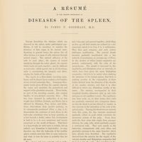 0103 - Page 35 - A résumé of the present knowledge of Disease of the spleen by James.F. Goodhart, M. D
-
 0105 - Page 37 - Capsule . Trabecule
-
 0107 - Page 39 - Vascular changes
-
 0108 - Page 40 - Arteries
-
 0111 - Page 43 - Lesions of the pulp and lymphoid tissues
-
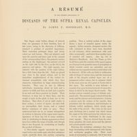 0117 - Page 49 - A résumé of the present knowledge of diseases of the supra renal capsules by James F. Goodhart, M.D
-
 0123 - Page sans numérotation - An atlas of illustrations of pathology Fasciculus III Diseases of the liver
-
 0126 - Page sans numérotation - Plate XI / Lymphadenoma of the liver
-
 0130 - Page sans numérotation - Plate XII / Fig. 1. Dilatation of the bile ducts in the liver from the pressure of a gallstone in the cystic duct. Fig. 2. Cancer of the liver with dilatation of the ducts and staining of the hepatic tissue
-
 0134 - Page sans numérotation - Plate XIII / Syphilitic cirrhosis of the liver
-
 0138 - Page sans numérotation - Plate XIV / Fig. 1. Red atrophy with acute yellow atrophy of the liver. Fig. 2. Microscopical appearances of the yellow swollen parts of the liver (acute yellow atrophy). Fig. 3. Microscopical appearances of red atrophy of the liver
-
 0142 - Page sans numérotation - Plate XV / Fig. 1. Lardaceous liver. Fig. 2. Lardaceous liver showing the iodine reaction
-
 0146 - Page sans numérotation - Plate XVI / Fig. 1. Cancer of the liver. Fig. 2. Nutmeg liver. Chronic congestion and atrophy of the liver from mitral disease
-
 0149 - Page 1 - Appendix / Plate XI. Lymphadenoma of the liver
-
 0150 - Page 2 - Plate XII / Fig. 1. Dilatation of the bile ducts
-
 0151 - Page 3 - Fig. 2. Cancer of the liver. Plate XIII .Syphilitic cirrhosis of the liver. Plate XIV. Fig. 1. Acute yellow and red atrophy of the liver. Figs. 2 and 3 represent the microscopical appearances found in the specimen depicted in fig. 1
-
 0152 - Page 4 - Plate XIV
-
 0153 - Page 5 - Plate XV
-
 0154 - Page 6 - Plate XVI / Fig. 1. Cancer of the liver
-
 0155 - Page 7 - Plate XVI / Fig. 2. Nutmeg liver
-
 0157 - Page sans numérotation - An atlas of illustrations of pathology Fasciculus IV diseases of the liver including one figure of spleen
-
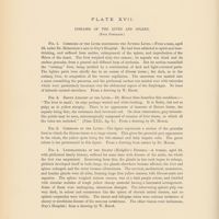 0160 - Page sans numérotation - Plate XVII / diseases of the liver and spleen . Fig. 1. Cirrhosis of the liver resembling the Nutmeg liver Fig. 2. Brown Atrophy of the liver. Fig. 3. Cirrhosis of the liver. Fig. 4. Lymphadenoma of the spleen ( hodgkin's Disease)
-
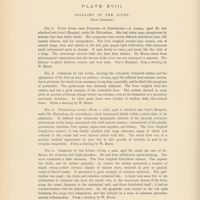 0164 - Page sans numérotation - Plate XVIII / Fig. 1. Fatty liver from poisoning by phosphorus. Fig. 2. Cirrhosis of the liver. Fig. 3. Tubercular liver. Fig. 4. Cirrhosis of the liver
-
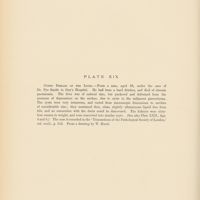 0168 - Page sans numérotation - Plate XIX / Cystic disease of the liver
-
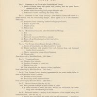
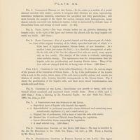 0171 - Page sans numérotation - Plate XX / Fig. 1. Lardaceous disease of the liver. Fig. 2. Fatty liver. Fig. 3. Early cirrhosis. Figs. 4 & 5 Cirrhosis of the liver (after Hamilton). Fig. 6. Cirrhosis of the liver. Fig. 7. A vegetation from the surface of the liver. Fig. 8. Spindle-cell sarcoma of the liver. Fig. 9. Disseminated growths of fibrous nature in the liver
-
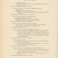
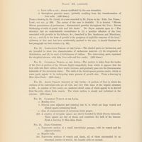 0172 - Page sans numérotation - Plate XX / Fig. 10. Lardaceous disease of the liver. Fig. 11. Cavernous tumour in the liver. Fig. 12. Acute yellow atrophy of the liver. Fig. 13. Cavernous tumour in the liver. Fig. 14 Early Cirrhosis. Fig. 15. Columnar epithelioma of the liver
-
 0175 - Page sans numérotation - Plate XXI / Fig. 1. Cirrhosis of the liver. Fig. 2. Cirrhosis of the liver showing a cross-section of ducts and vessels in a portal channel, with the surrounding changes. Fig. 3. monolobular cirrhosis (after Dreschfeld and Young). Fig. 4. The nutmeg liver (Ramose atrophy of Moxon). Fig. 5. Tubercular liver. Fig. 6. The nutmeg liver, showing appearances in the portal canals similar to those of the so-called biliary cirrhosis. Fig. 7. Miliary gummata. Fig. 8. Idiopathic anaemia. Atrophy in the distribution of the hepatic artery and intrabular vein
-
 0176 - Page sans numérotation - Plate XXI / Figs. 9 &10 Cancer of the bile ducts. Fig. 11. Cancer spreading from the biliary ducts. Fig. 12. Early gummatous infiltration of the liver. Fig. 13 "Common " cirrhosis ( "Multilobular" Cirrhosis). Fig. 14. Tubercular Liver. Fig. 15. Idiopathic Anaemia
-
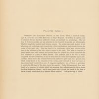
 0179 - Page sans numérotation - Plate XXII / Fig. 1. "Pericellular" Cirrhosis. Fig. 2. Cirrhosis of the liver. Fig. 3. Nutmeg liver,showing cirrhotic changes. Fig. 4. Cystic liver to show the changes in the portal canals. Fig. 5. Cystic liver. Fig. 6. Early cancer of the liver. Fig. 7. Extreme tubercular disease of liver. Fig. 8. Brown atrophy of the liver
-
 0180 - Page sans numérotation - Plate XXII / Fig. 9. Extreme tubercular disease of the liver. Fig. 10. Myxoedematous liver. Figs. 11,12 & 13 "Contracting scirrhus of the liver simulating cirrhosis". Fig. 11. Isolated cells from nodules of cancer, large and not at all unlike the gland cells. Fig. 12. Part of the infiltrating growth. Fig. 13. Shows the cells from a nodule of the growth in position. Figs. 14, 15 & 16 Varieties of cell vacuolation and proliferation, illustrating the stages of cancer in the liver. Fig. 17. Primary adenoma of the liver. Fig. 18. Leukaemic liver. Fig. 19. Primary adenoma of the liver
-
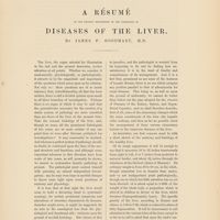 0183 - Page 1 - A résumé of our present knowledge of the pathology of diseases of the liver by james F. Goodhart, M.D
-
 0187 - Page 5 - Morbid Histology / Hypertrophy
-
 0188 - Page 6 - Fatty infiltration. Atrophy. Fatty degeneration. Granular degeneration
-
 0189 - Page 7 - Lardaceous degeneration. Proliferation of the hepatic cells
-
 0190 - Page 8 - Connective Tissue and its canals biliary and vascular
-
 0193 - Page 11 - Diseases of the liver and its capsule. Hypertrophy
-
 0194 - Page 12 - Atrophy / Simple atrophy. Atrophy from pressure
-
 0195 - Page 13 - Brown or chronic Atrophy
-
 0196 - Page 14 - Acute yellow atrophy / Morbid anatomy. Histology. Yellow parts
-
 0202 - Page 20 - Phosphorus poisoning
-
 0204 - Page 22 - The fatty liver
-
 0205 - Page 23 - Lardaceous disease
-
 0206 - Page 24 - Histology
-
 0207 - Page 25 - Diseases due to circulatory disturbance / Oedema.Hyperoemia.The nutmeg liver
-
 0208 - Page 26 - General appearances. Histology
-
 0209 - Page 27 - Anaemia
-
 0210 - Page 28 - Diseases of inflammatory nature / Perihepatitis
-
 0211 - Page 29 - Hepatic abscess
-
 0213 - Page 31 - Cirrhosis
-
 0215 - Page 33 - Histology. Minute structure of the cirrhotic material
-
 0220 - Page 38 - Syphilitic hepatitis / Congenital syphilitic hepatitis
-
 0221 - Page 39 - Syphilitic hepatitis in the adult
-
 0222 - Page 40 - The pigmented liver / New growths. Angeimata or Cavernous tumours
-
 0225 - Page 43 - Adenoma and primary cancer
-
 0226 - Page 44 - Tubular Adenoma. Primary Cancer
-
 0227 - Page 45 - Lymphatic Tumours
-
 0229 - Page 47 - Cysts
-
 0230 - Page 48 - Hydatid disease
-
 0231 - Page 49 - Malformations. Injuries . Disease of the portal vein
-
 0234 - Page 52 - Appendix / The liver of phosphorus poisoning
-
 0235 - Page sans numérotation - An atlas of illustrations of pathology Fasiculus V diseases of the liver (chiefly of the gall bladder and larger bile ducts)
-
 0238 - Page sans numérotation - Plate XXIII / Syphilitic and lardaceous disease of the liver
-
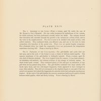 0242 - Page sans numérotation - Plate XXIV / Fig. 1. Abscesses in the liver. Fig. 2. Papilloma of the Gall- Bladder
-
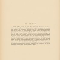 0246 - Page sans numérotation - Plate XXV / Cancer of gall-bladder and liver.Gall-stones , with obstruction and dilatation of the cystic duct
-
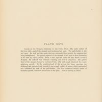 0250 - Page sans numérotation - Plate XXVI / Cancer of the stomach extending to the cystic duct
-
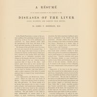 0253 - Page sans numérotation - A résumé of our present knowledge of the diseases of the liver (Gall bladder and larger bile ducts) by James F. Goodhart, M.D
-
 0259 - Page sans numérotation - An atlas of illustrations of pathology Fasciculus VI Hydatid disease of liver urinary calculi
-
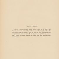 0262 - Page sans numérotation - Plate XXVII / Part of a liver containing several hydatid cysts
-
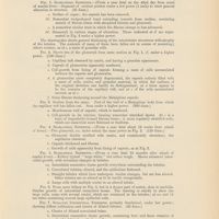
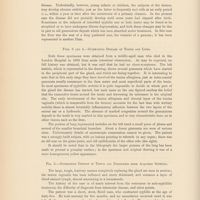
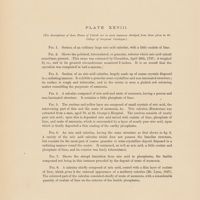 0266 - Page sans numérotation - Plate XXVIII / Fig. 1. Section of an ordinary large uric acid calculus, with a little oxalate of lime. Fig. 2. Shows the polished, tuberculated, or granular, exterior which uric acid calculi sometimes presents. Fig. 3. Section of an uric acid calculus, largely made up of coarse crystale disposed in a radiating manner . Fig. 4. A calculus composed of uric acid and urate of ammonia, having a porous and non-laminated structure. Fig. 5. The nucleus and yellow layer are composed of small crystals of uric acid, the intervening part of this and the urate of ammonia, &c. Fig. 6. An uric acid calculus, having the same structure as that shown in fig. 3. Fig. 7. Shows the abrupt transition from uric acid to phosphates, the fusible compound not being in this instance preceded by the deposit of urate of ammonia. Fig. 8. A calculus chiefly composed of uric acid, coated with a thin layer of oxalate of lime,which gives it the external appearance of a mulberry calculus
-
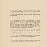 0270 - Page sans numérotation - Plate XXIX / Fig. 1. Section of an uric acid calculus from the kidney. Fig. 2. External view of the same. Fig. 3. Section of a large calculus made up of three distinct uric and acid stones, united by earhy phosphates. Fig. 4. Section of a singular but not very uncommon form of calculus, the centre consisting of uric acid, the pinkish gray part of urate of ammonia and uric acid, the white layer at the sides of phosphate of lime. Fig. 5 & 6 Exterior and section of a cystic oxide calculus. The confusedly crystalline structureis well seen in fig.6, wich represents a section and the minutely tubercular appearance of its exterior in fig. 5 . Fig. 7. A calculus almost entirely made up of uric acid and urate of ammonia deposited on a piece of steel apparently the end of a stilet. Fig. 8. The nucleus and exterior consist of uric acid and urate of ammonia,the intermediate layer of oxalate of lime. Fig. 9. A calculus chiefly made up of oxalate of lime, having a nucleus of impure urate of ammonia, and a white layer chiefly consisting of phosphate of lime Figs. 10 & 11 Section and exterior of small uric acid calculus, thinly coated with urate of ammonia. Fig. 12. Shows the crystalline centre and laminated structure of a very characteristic specimen of pisiform, uric acid concretion
-
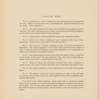 0274 - Page sans numérotation - Plate XXX / Fig. 1. A gray layer of urate of ammonia is seen occuring between the deposits of uric acid. Fig. 2. The yellow nucleus is uric acid; it is surrounded by a gray layer of urate of ammonia; then follow alternating layers of urate of ammonia and the fusible phosphates; on the outside is an irregular deposit of triple phosphate. Fig. 3. Represens the ordinary appearances of the urate of ammonia calculus. Fig. 4. Section of a calculus consisting of urate of ammonia with urate of lime. Fig. 5. The section of a calculus, consisting of urate of ammonia surrounded by phosphates. Fig. 6. The nucleus consists of urate of ammonia with oxalate of lime; around this is white oxalate of lime mixed with some phosphate of lime; the whole is coated with pure oxalate of lime, upon wich is a partial deposit of uric acid. Fig. 7 shows the abrupt and well-defined transition from urate of ammonia to oxalate of lime; the exterior is thinly coated with a layer of phosphate and oxalate of lime. Fig. 8. The ordinary phosphate of lime calculi found in the cells of the prostate gland. Fig. 9. The ordinary " hemp-seed" calculi, consistingof oxalate of lime and urate of ammonia. Figs. 10, 11 & 12 Earthy phosphate calculi taken from a cyst in the prostate gland
-
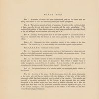 0278 - Page sans numérotation - Plate XXXI / Fig. 1. A calculus, of which the inner tuberculated part and the outer layer are nearly pure oxalate of lime the intervening white part fusible phosphates. Fig. 2. The nucleus consists of urate of ammonia. Fig. 3. Calculus showing a thin layer of uric acid deposited on a mass of oxalate of lime. Figs.4 & 5 Represent the white crystalline. Figs. 6,7,8,9, & 10 . Carbonate of lime calculi . Fig. 11 Represents the central portion, and one of the fragments of a large uric acid calculus. Fig. 12. the centre consists of urate of ammonia surrounded by oxalate of lime, divided into two by a thin layer of phosphates. Fig. 13. The external surface of a calculus which has under gone partial solution in the bladder. Fig. 14. A section of the same. In this drawing are shown the abrupt termination of the outer uric acid layers,together with the thickness of the layer of the fusible compound, which has been deposited over the whole of its exterior
-
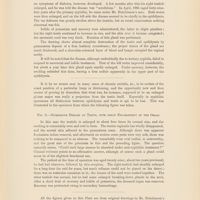
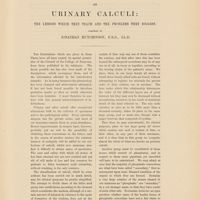 0281 - Page sans numérotation - On Urinary calculi : the lessons wich they teach and the problems they suggest. Compiled by Jonathan Hutchinson, F.R.S., LL.D
-
 0282 - Page 2 - Salts which enter into the composition of calculi / Uric Acid. The Uric Oxide Calculus
-
 0283 - Page 3 - Urates. Oxalate of lime
-
 0284 - Page 4 - Cystine or cystic acid. Xanthine or Xanthic Oxide. Carbonateof lime
-
 0285 - Page 5 - The triple phosphate. Phosphatic calculi and the " phosphatic diathesis"
-
 0290 - Page 10 - Cystine Calculi
-
 0294 - Page 14 - Mixed calculi. On the nuclei of stones
-
 0296 - Page 16 - Calculi containing Indigo. On the influence of sex in relation to stone
-
 0297 - Page 17 - On the possible explanations of local prevalence of Calculus
-
 0304 - Page 24 - On Urinary in the lower Animalis
-
 0305 - Page 25 - On Calculi of unusual size. Accretions on foreign bodies
-
 0307 - Page 27 - Concluding remarks
-
 0311 - Page sans numérotation - An atlas of illustrations of pathology fasciculus VII Urinary calculi and gall-stones. Enlargement of the prostate gland. Enlargement of prostate, urinary calculi &c. Osteitis deformans ( pagets disease) with descriptive letterpress
-
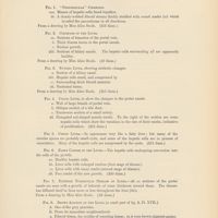
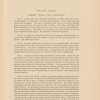 0313 - Page sans numérotation - Plate XXXII / Urinary calculi andgall-stones. Fig. 1. A bone bodkin-case, with thread attached to its middle, which was removed from the blader of a lady about a week after its introduction. Fig. 2. A portion of sealing-wax doubled up and encrusted with phosphates. Fig. 3. A urinary calculus, seen in section, of a very remarkable shape. Fig. 4. A calculus concretion which had formed on a bit of straw. Fig. 5,6,7,8,9, 10,11,12 A series of gall-stones, which were passed per anum
-
 0314 - Page sans numérotation - Plate XXXII / Fig. 13. Shows the size and shape of calculus. Fig. 14. A large gall-stone. Figs. 15., 16 & 17 incomplete obstruction of the bowels
-
 0316 - Page sans numérotation - Plate XXXII / general remarks
-
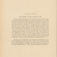 0320 - Page sans numérotation - Plate XXXIII / Enlargement of the prostate gland. This plate shows the relations assumed by the bladder and rectum in a case of enormous enlargement of the prostate
-
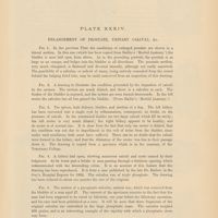 0323 - Page sans numérotation - Plate XXXIV / Enlargement of prostate, urinary calculi, & c. Fig. 1. In the previous Plate the conditions of enlarged prostate are shown in a lateral section. Fig. 2. A drawing to illustrate the condition presented by the impaction of calculi by the ureters. Fig. 3. The spleen both kidneys, bladder and urethra of a dog. Fig. 4. A kidney laid open,showing numerous calculi and cysts caused by their lodgment. Fig. 5. The section of a phosphatic calculus. Fig 6. A very large calculus
-
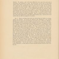 0324 - Page sans numérotation - Plate XXXIV / Fig. 7 shows a yet larger stone which was removed from the bladder of a woman withouit any operation
-
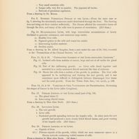
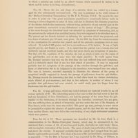
 0328 - Page sans numérotation - Plate XXXV / osteitis deformans. Figs. 1,2 and 3 show his general contour six months before death ; the position in wich his head was carried, and the bending and enlargement of the bones of the left lower extremity, are well seen. Fig 4. we have a section of his skull,showing its great enlargement by the external deposit of loose porous bone. Figs. 5 and 6 show sections of the upper and lower parts of the femur. Fig. 7. illustrates the way in wich the fibula was bent
-
 0331 - Page sans numérotation - An atlas of illustrations of PathologyFasciculus VIII Diseases of brain and spinal cord
-
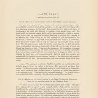 0333 - Page sans numérotation - Plate XXXVI./ Fig. 1. Hydatid in the posterior cornu of the right lateral ventricle. Fig. 2. Abscess on the under surface of the right cerebellar hemisphere close to the petrous portion of the temporal bone
-
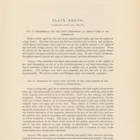 0337 - Page sans numérotation - Plate XXXVII / Fig. 1.Haemorrhage into the right hemisphere and median lobe of the cerebellum. Fig. 2. Tubercles of various sizes situated on the upper surface of the cerebellar hemispheres
-
 0338 - Page sans numérotation - Plate XXXVII / Fig . 3. A tuberculous tumour situated between the left side of the pons Varolii, the medulla oblongata,and the adjacent surface of the left cerebellar hemisphere
-
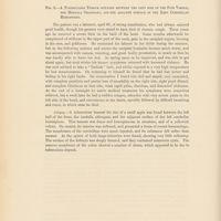
 0341 - Page sans numérotation - Plate XXXVIII / Fig. 1. A severely crushed Spinal Cord. Fig. 2. The cervical spinal cord of a man who had died under almost precisely similar conditions to those specified in the preceding case
-
 0342 - Page sans numérotation - Plate XXXVIII / Fig. 3. Haemorrhage external to the vertebral theca
-
 0346 - Page sans numérotation - Plate XXXIX / Figs. 1, 2, 3. A Tuberculous tumour non the spinal dura mater
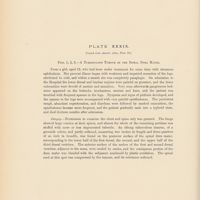
-
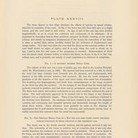
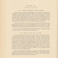 0350 - Page sans numérotation - Plate XL / Fig.1 Cartilaginous deposits on the spinal arachnoid. Fig. 2. Myelitis after concussion of the spine
-
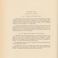 0354 - Page sans numérotation - Plate XLI / Fig. 1 Tubercle in pia mater of cord. Fig. 2. A fibrous tumour lodged in the cauda equina
-
 0357 - Page sans numérotation - An atlas of illustrations of pathology fasciculus IX diseases of the testis (part I)
-
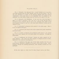 0360 - Page sans numérotation - Plate XLII /Fig. 1. Hydrocele of the Spermatic Cord. Fig. 2.Hydocele of th Epididymis. Fig . 3 Pedunculated cartilaginious body attached to the globus major. Fig. 4. A calcareous plate formed in the tunica vaginalis. Fig. 5 A numberof calcareous and .cartilaginous bodies formed in the visceral layer of the tunica vaginalis and on the globus major of the epididymis. Fig. 6. A multicular cyst deveioped "between the tunica vaginalis and the tunica albuginea" Fig. 7. A hydroceleof the uppert part of the tunica vaginalis, the lower part having become obliterated by adhesion to the testicle,wich is seen insection
-
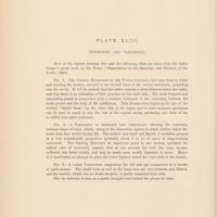 0364 - Page sans numérotation - Plate XLIII / Hydocele et varicocele. Fig. 1. The common hydrocele of the tunica vaginalis laid open from in front, and showing the testicle, covered by its visceral layer of the serous membrane, projecting into the cavity. Fig. 2. A varicocele of moderate size unravelled,showing the extremely tortuous loops of veins,wich,owing to the dissection appear to reach further below the testis than they would during life.Fig. 3. A large varicocele, suggesting the old and apt comparison to a bundle of earth-worms
-
 0368 - Page sans numérotation - Plate XLIV / Fig. 1 Undescended and atrophied testis. Fig. 2. Atrophy ( extreme) of one testicle and epididymis. Fig . 3 Cystic disease ( ? Sarcoma) of the testis
-
 0371 - Page sans numérotation - Plate XLV / Syphilis of the testicle. Fig. 1. Breaking down gumma in the testis. Fig. 2. Gumma of the testis due to inherited syphilis
-
 0372 - Page sans numérotation - Plate XLV / Figs. 3 and 4 Gummatous disease of testis and lung. Fig. 5. Gummatous deposit in testis and epididymis from acquired syphilis
-
 0373 - Page sans numérotation - Plate XLV / Fig. 6. Gummatous disease of testis, with great enlargement of the organ
-
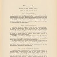 0377 - Page sans numérotation - Plate XLVI / Tumour of the testicle. Gumma of the testicle. Fig. 1. Medullary cancer. Fig. 2. Cystic chondro-sarcoma. Fig. 3 Gumma of testicle, with hydrocele
-
 0378 - Page sans numérotation - Plate XLVI / Fig. 4. A sarcoma involving the whole testicle, and spreading up the spermatic cord
-
 0381 - Page sans numérotation - An atlas of illustrations of pathology fasciculus X diseases of the testis ( part II)
-
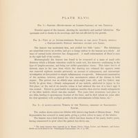 0384 - Page sans numérotation - Plate XLVII / Fig. 1. Sarcoma (round-celled or lympho-sarcoma) of the testicle. Fig. 2. View of an antero-posterior section of the above tumour, showing a greyish-brow surface obscurely divided into lobes. Fig 3. A slowly- growing tumour of the testicle, probably of sarcomatous nature
-
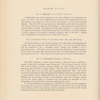 0388 - Page sans numérotation - Plate XLVIII / Fig. 1. Hydrocele of tunica albuginea. Fig. 2. Malignant tumour of the testis from a boy aged two years. Fig. 3. Haemorrhagic sarcoma of testicle
-
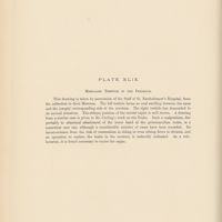 0392 - Page sans numérotation - Plate XLIX / Misplaced testicle in the perineum
-
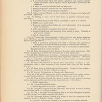
 0395 - Page 1 - The pathology of the testis / I Hydrocele of the Cord (Plate XLII. Fig. 1.)
-
 0397 - Page 3 - II Hydrocele of the epididymis. (Plate XLII. Fig. 2.) / 1 Subserous or lenticular cysts. 2. The large cysts of the epididymis ( spermatic cysts of the French writers, encysted or parenchymatous hydrocele of the epididymis, Curling )
-
 0398 - Page 4 - III Hydrocele of the tunica vaginalis (Plate XLIII. Fig. 1)
-
 0402 - Page 8 - IV Cartilaginous and calcareous bodies in the tunica vaginalis, &c. (Plate XLII. Figs. 3,4 and 5)
-
 0403 - Page 9 - V. Varicocele. (Plate XLIII. Figs. 2 and 3) / 1 The proportion of cases of varicocele in which the testicle is more or les atrophic is variably estimated by different obsevers. 2 Relaxation of the scrotum , &c
-
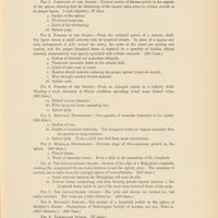
 0405 - Page 11 - VI retention and malposition of the testicles ( Plate XLIV.Fig.1; and plate XLIX)
-
 0406 - Page 12 - Torsion of the cord with haemorrhage into, atrophy or gangrene of the testis
-
 0408 - Page 14 - Development of tumours
-
 0409 - Page 15 - On imperfect descent in relation to the function of the testis
-
 0411 - Page 17 - Tubercular disease of the testis
-
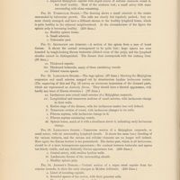
 0413 - Page 19 - VII Syphilitic Orchitis (Plates XLV and XLVI., Fig 3. )
-
 0415 - Page 21 - The complications of Syphilitic Orchitis
-
 0416 - Page 22 - The co-existence of Syphilitic disease of the lungs and testes
-
 0417 - Page 23 - IX Tumours of the testicle / Carcinoma
-
 0418 - Page 24 - Sarcoma. Haemorrhagic sarcoma. Lympho sarcoma
-
 0419 - Page 25 - Cystic disease of the testicle (Plates XLIV. Fig.3. and XLVI. Fig. 2.)
-
 0421 - Page 27 - Growths secondary to tumours of the testis
-
 0422 - Page 28 - Malignant tumours starting in a retained testis
-
 0423 - Page 29 - Malignant tumours of the testis in early life X Haematocele
-
 0424 - Page 30 - XI Atrophy of the testis
-
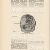
 0428 - Page sans numérotation - The plates from I . To VI inclusive, have reference to the case of John M'Cann, related at page 21./ Plate I External appearances of the body
-
 0432 - Page sans numérotation - Plate II Neuroma of the left sciatic nerve,ten inches in its transverse and eleven in its vertical diameter
-
 0436 - Page sans numérotation - Plate III / Fig. 1. The right sciatic nerve, presenting at its superior part a large tumour,five inches in length. Fig 2 The anterior and posterior tabial and the external saphenus nerves of the left side. The large tumour occupied the lower part of the popliteal space
-
 0440 - Page sans numérotation - Plate IV / Fig.1. The right anterior crural nerve and its branches from the lumbar plexus to the knee. Fig 2. The left anterior crural and its principal branches crowded with tumours, upon the surface of many of which nervous filaments are seen
-
 0444 - Page sans numérotation - Plate V / Fig 1. The nerves of the right upper extremity. Fig. 2. Nerves of the left upper extremity
-
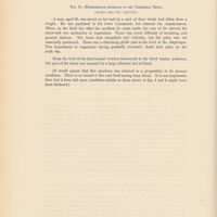
 0448 - Page sans numérotation - Plate VI / Fig. 1. The right pneumogastric nerve immensely enlarged, and having connected with its cervical portion a neuroma of great size. Fig. 2. The neuroma represented in Fig. 1. , laid open, and in part detached from its capsule. Fig. 3. This figure represents the fourth,fifth and sixth intercostal nerves. Fig. 4. Neuromatous tumours upon the under surface of the tongue connected with the branches of the left hypoglossal nerve. Fig. 5. Small neuromatous tumours connected with the left phrenic nerve in the thorax.Fig. 6. The cervical portion of the left pneumogastric nerve, greatly enlarged, and covered with tumours. Fig. 7. Neuromatous tumours which existed in the pelvis connected with anterior branches of the sacral nerves. Fig. 8, 9, 10, 11 these figures represent neuromatous tumours upon the delicate fibrillae of different nerves
-
 0452 - Page sans numérotation - The plates from VII. To XIII. Inclusive, illustrate the morbid appearances observed in the case of Michael Lawlor, described at page 26 / Plate VII The front of the abdomen and of the lower part of the thorax, covered with a vast multitude of small superficial, neuromatous tumours
-
 0456 - Page sans numérotation - Plate VIII / Fig. 1. The right upper extremity, in wich chains of tumours mark the course of the cutaneous nerves,rendering them visible through the integuments. Fig. 2. The nerves of the right arm and fore-arm studded with tumours of various sizes throughout their entire course, from the brachial plexus to the hand
-
 0460 - Page sans numérotation - Plate IX / Fig. 1. The right anterior crural nerve and its branches, together with a portion of the lumbar plexus. Fig. 2. Nerves of the left upper extremity, greatly enlarged,and presenting numerous tumours
-
 0464 - Page sans numérotation - Plate X / Fig. 1. This figure represents the oesophagus and the pneumogastric nerves in the thoracic division of their course. Fig. 2.The popliteal and tibial nerves of the right side. Fig. 3. The branches of the left anterior crural nerve. Figs. 4. 5. The portions of phrenic nerves which correspond to the pericardium, presenting severaol small oblong tumours
-
 0468 - Page sans numérotation - Plate XI / The right sciatic nerve,from the sacral plexus to the knee. The large tumours is connected with the plexus; it filled the true pelvis
-
 0472 - Page sans numérotation - Plate XII / Fig.1. The left sciatic nerve covered with tumours.Fig. 2. A posterior view of the tumour represented in Fig. 1, showing the separation of the fibres of the nerve from each other. Fig. 3. This figure exhibits several small cavities in the interior of the tumours, and likewise shows the two capsules by wich it was invested, separated from each other
-
 0476 - Page sans numérotation - Plate XIII / Fig. 1. Neuroma connected with the external branch of the median nerve distributed to the index finger. The relation of the tumour to the nerve is seen in Fig. 2. Fig. 3. This figure, copied from the work of Cruveilhier, shows a neuroma connected with the internal branch of the median nerve distributed to the index finger. Fig. 4. This figure (also copied from the work of Cruveilhier represents a spheroidal neuromatous tumour, connected with the musculo-spinal nerve at the bend of the elbow. Fig. 5. Neuroma developed in the centre of the popliteal nerve. Fig. 6. Neuroma of the ulnar nerve. Fig. 7. A serous cyst, developed among the branches of the median nerve distributed to the thumb. Fig. 8. Neuroma of the ulnar nerve; case recorded by Cheselden. Fig 9. Neuroma of the lower part of the sciatic nerve. Fig. 10. A portion of the brachial plexus, showing remarkable fusiform enlargements of the roots of the median nerve. Fig. 11. This figure, copied from the work of Cruveilhier, represents enlargements of nerves, somewhat resembling those delineated in Fig. 10. Fig. 12. Neuroma of the Gasserian ganglion
-
 0480 - Page sans numérotation - Plate XIV / Fig. 1. This figure represents the appearances of the extremities of the nerves in a case where the fore-arm had been amputed near its centre. Figs. 2, 3. The sciatic nerve removed from a stamp. Fig. 4. The détails of the dissection of a stump, after amputation of the arm near its centre, are represented in this Figure. Figs. 5, 6. Fig. 5. represents a tumour connected with the extremities of the nerves of the brachial plexus, in a case in wich amputation at the shoulder-joint was performed by Baron Larrey. The termination of the fibres of the nerves in the interior of the tumour is seen in FIg. 6. Fig 7. A portion of the median nerve in the fore-arm, removed from a stump. Figs. 8, 9.The sciatic nerves of stumps. Fig 10. The sciatic nerve of stump. Fig. 11. Traumatic neuromatous tumour of the median nerve
-
 0484 - Page sans numérotation - Plate XV / Fig. 1. It represents the disease generally known by the name of the "Painful subcutaneous tubercle ". Fig. 2. The left semilunar ganglion greatly enlarged. Fig 3. The right semilunar ganglion in the same case. Fig. 4. Hypertrophy of the cervical ganglia of the sympathetic nerve. Fig. 5 represents the inferior and Fig . 6. The superior cervical ganglion in the case of Erasmus Saulich,detailled at p.20. Figs. 7,8, 9 These figures represent the tumours upon the extremities of the nerves in the case of John Byrne, recorded at p.36. Fig.10 . A magnified view of the fibres in the interior of tumour which formed upon the extremity of the nerf of a stump. Fig. 11. This figure exhibitis the appearance which a portion of the neuroma of the Gasserian ganglion, described at p.31, presented when examined by the aid of the microscope. Fig.12. exhibits the cellular structure of a portion of an idiopathic neuroma. Fig. 13. represent the Pacinian corpuscles connected with the digital branches of the median nerve.Fig. 14. Atrophy of the right optic nerve and the left optic tract,in a case where right eye had been destroyed by small-pox many years previous to death
-
 0487 - Page 1 - A treatise on the pathology, diagnosis, and treatment of neuroma
-
 0489 - Page 3 - Prefatory note
-
 0491 - Page 5 - A treatise on the pathology, diagnosis, and treatment of neuroma
-
 0492 - Page 6 - General characters and symptoms of idiopathic neuroma
-
 0499 - Page 13 - The treatment of neuromata
-
 0501 - Page 15 - Neuroma of the median nerve. Neuroma of the internal cutaneous nerve of the arm. Neuroma of the axillary plexus
-
 0504 - Page 18 - General development of Neuromatous tumours
-
 0506 - Page 20 - Autopsy
-
 0510 - Page 24 - Hypoglossal nerve
-
 0513 - Page 27 - Nerves of the right lower extremity. Nerves of the left lower extremity
-
 0514 - Page 28 - Nerves of the right upper extremity. Nerves of the left upper extremity. Pneumogastric nerves
-
 0515 - Page 29 - Neuroma of the ganglionic system
-
 0516 - Page 30 - Co incidence of multiple neuromata with mental defects
-
 0517 - Page 31 - Solitary neuroma of gasserian ganglion. Traumatic neuroma
-
 0518 - Page 32 - Traumatic neuroma of the median nerve. Traumatic neuroma of the ulnar nerve
-
 0519 - Page 33 - Traumatic neuroma of the radial nerve
-
 0521 - Page 35 - Neuroma succeeding to amputations
-
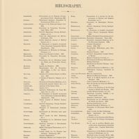 0525 - Page sans numérotation - Bibliography
-
 0527 - Page sans numérotation - An atlas of illustrations of pathology fasciculus XII infective disease of the lymphatic system : lymph-adenoma, or Hodgkin's malady
-
 0529 - Page 1 - Infective disease of the lymphatic system. Extract from DR. Hodgkin's paper
-
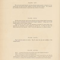 0532 - Page sans numérotation - Plate LXV The neck and armpit dissected,showing enlarged glands grouped in bunches of very various sizes, and with but little tendeney to become adherent together. Plate LXVI The iliac fossa and upper part of thigh dissected, showing enlarged glands surrounding the iliac vessels, and passing with them under Poupart's ligament. Figs II. , III., IV. , and V. show the appearances presented by single glauds, more especially the vascularity of their capsules,&c. Plate LXVII Fig. I. shows the spleen in section. Fig. II shows the size and condition of the enlarged spleen. Plate LXVIII Fig. I. A section through the liver and the large mass of glands beneath it. Fig II Front viewof the liver and gland-mass
-
![0545 - Page sans numérotation - [Contreplat]](https://numerabilis.u-paris.fr/iiif/2/bibnum:01866:0545/square/200,/0/default.jpg) 0545 - Page sans numérotation - [Contreplat]
0545 - Page sans numérotation - [Contreplat]
-
![0546 - Page sans numérotation - [Plat]](https://numerabilis.u-paris.fr/iiif/2/bibnum:01866:0546/square/200,/0/default.jpg) 0546 - Page sans numérotation - [Plat]
0546 - Page sans numérotation - [Plat]
- Autre consultation
- Consulter en PDF