
Myotomia reformata : or an anatomical treatise on the muscles of the human body. Illustrated...
- Collections
- Anatomie
- DESCRIPTION
- TABLE DES MATIÈRES
- VOIR PLUS
- Identifiant
- ark:/13685/01892
- Titre
- Myotomia reformata : or an anatomical treatise on the muscles of the human body. Illustrated with figures after the life. By the late Mr. William Cowper, Surgeon, and Fellow of the Royal Society. To which is prefix'd an Introduction concerning muscular motion
- Créateur
- Cowper, William
- Date
- 1724
- Éditeur
- London : printed for Robert Knaplock, and William and John Innys, in St. Paul's Church-Yard; and Jacob Tonson, in the Strand
- Siècle
- 18e siècle
- Format
- Nombre de vues : 434
- Source
- Université Paris Cité. BIU Santé Médecine, inv. 1892
- Date de mise en ligne
- 25 octobre 2016
- Propriétaire
- Université Paris Cité. BIU Santé Médecine
- Licence
- Licence Ouverte
- Table des matières
-
![0001 - Page sans numérotation - [Plat]](https://numerabilis.u-paris.fr/iiif/2/bibnum:01892:0001/square/200,/0/default.jpg) 0001 - Page sans numérotation - [Plat]
0001 - Page sans numérotation - [Plat]
-
![0002 - Page sans numérotation - [Contreplat]](https://numerabilis.u-paris.fr/iiif/2/bibnum:01892:0002/square/200,/0/default.jpg) 0002 - Page sans numérotation - [Contreplat]
0002 - Page sans numérotation - [Contreplat]
-
![0003 - Page sans numérotation - [Page de garde]](https://numerabilis.u-paris.fr/iiif/2/bibnum:01892:0003/square/200,/0/default.jpg) 0003 - Page sans numérotation - [Page de garde]
0003 - Page sans numérotation - [Page de garde]
-
![0007 - Page sans numérotation - [Frontispice]](https://numerabilis.u-paris.fr/iiif/2/bibnum:01892:0007/square/200,/0/default.jpg) 0007 - Page sans numérotation - [Frontispice]
0007 - Page sans numérotation - [Frontispice]
-
![0009 - Page sans numérotation - [Page de titre]](https://numerabilis.u-paris.fr/iiif/2/bibnum:01892:0009/square/200,/0/default.jpg) 0009 - Page sans numérotation - [Page de titre]
0009 - Page sans numérotation - [Page de titre]
-
 0011 - Page sans numérotation - Advertisement
-
 0013 - Page sans numérotation - The Preface
-
![0022 - Page sans numérotation - Syllabus Musculorum eâ serie qua in ordinariis Corporis Humani [anatomais] dissecantur](https://numerabilis.u-paris.fr/iiif/2/bibnum:01892:0022/square/200,/0/default.jpg) 0022 - Page sans numérotation - Syllabus Musculorum eâ serie qua in ordinariis Corporis Humani [anatomais] dissecantur
0022 - Page sans numérotation - Syllabus Musculorum eâ serie qua in ordinariis Corporis Humani [anatomais] dissecantur
-
 0023 - Page LXXVI - Introduction
-
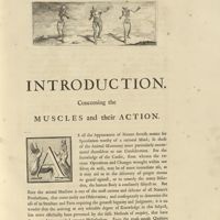 0025 - I - Introduction concerning the Muscles and their Action
-
 0029 - V - Part I
-
 0056 - Page XXXIV - Part II
-
 0057 - Page XXXV - Proposition I
-
 0059 - Page XXXVII - Proposition II
-
 0062 - Page XL - Proposition III
-
 0066 - Page XLIV - Proposition IV
-
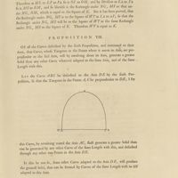
 0068 - Page XLVI - Proposition V
-
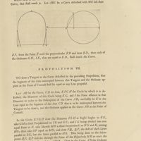
 0071 - Page XLIX - Corollary. Proposition VI
-
 0075 - Page LIII - Corollary I
-
 0076 - Page LIV - Corollary II / Corollary III
-
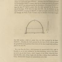
 0077 - Page LV - Corollary IV. Proposition VII
-
 0079 - Page LVII - Proposition VIII
-
 0082 - Page LX - Proposition IX
-
 0099 - Page LXXVII - Errata
-
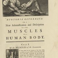 0101 - Page 1 - Myotomia reformata : or, a New Administration and Description of the Muscles of a Human Body. Chap. I. Of the Muscles of the Abdomen
-
 0102 - Page 2 - I. Obliquus descendens
-
 0103 - Page 3 - II. Obliquus ascendens
-
 0104 - Page 4 - III. Pyramidalis vel succenturiatus. IV. Rectus
-
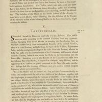 0105 - Page 5 - V. Transversalis
-
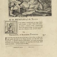 0107 - Page 7 - Chap. II. Of the Muscles of the Testes. VI. Cremaster, or Suspensor Testiculi
-
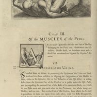 0108 - Page 8 - Chap. III. Of the Muscles of the Penis. VII. Accelerator urinae
-
 0109 - Page 9 - VIII. Erector Penis. IX. Transversalis Penis
-
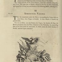 0110 - Page 10 - X. Erector Clitoridis. XI. Sphincter Vaginae
-
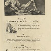 0111 - Page 11 - Chap. IV. Of the Muscles of the Bladder of urine. XII. Detrusor urinae. XIII. Sphincter vesicae
-
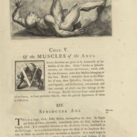 0113 - Page 13 - Chap. V. Of the Muscles of the Anus. XIV. Sphincter ani
-
 0114 - Page 14 - XV. Levator ani
-
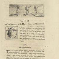 0115 - Page 15 - Chap. VI. Of the Muscles of the Hairy-Scalp and Forehead. XVI. Occipitalis
-
 0116 - Page 16 - XVII. Frontalis
-
 0117 - Page 17 - Chap. VII. Of the Muscles of the Cheeks and Lips. XVIII. Quadratus genae, or Quadratus colli, by some call'd Tetragonus, and by Galen, Platysma Myoides
-
 0118 - Page 18 - XIX. Buccinator. XX. Zygomaticus. XXI. Elevator labiorum
-
 0119 - Page 19 - XXII. Depressor labiorum. XXIII. Orbicularis labiorum. XXIV. Elevator labii superioris proprius. XXV. Depressor labii inferioris proprius
-
 0120 - Page 20 - XXVI. Elevator labii inferioris proprius
-
 0121 - Page 21 - Chap. VIII. Of the Muscles of the Eye-Lids. XXVII. Orbicularis palpebrarum
-
 0122 - Page 22 - XXVIII. Aperiens palpebram rectus
-
 0123 - Page 23 - Chap. IX. Of the Muscles of the Eye. XXIX. Obliquus superior or Trochlearis
-
 0124 - Page 24 - XXX. Obliquus inferior
-
 0125 - Page 25 - XXXI. Elevator oculi. XXXII. Depressor oculi. XXXIII. Adductor oculi. XXXIV. Abductor oculi
-
 0127 - Page 27 - Chap. X. Of the Muscles of the Nose. XXXV. Elevator alae nasi
-
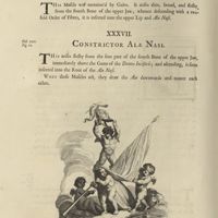 0128 - Page 28 - XXXVI. Dilatator alae nasi, & elevator labii superioris. XXXVII. Constrictor alae nasi
-
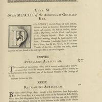 0129 - Page 29 - Chap. X. Of the Muscles of the Auricula or Outward Ear. XXXVIII. Attollens auriculam. XXXIX. Retrahens auriculam
-
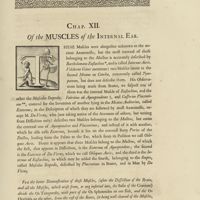 0131 - Page 31 - Chap. XII. Of the Muscles of the Internal Ear
-
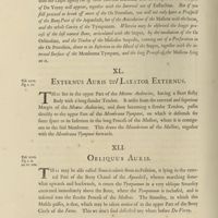 0132 - Page 32 - XL. Externus auris vel laxator externus. XLI. Obliquus auris
-
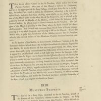 0133 - Page 33 - XLII. Internus auris. XLIII. Musculus Stapedis
-
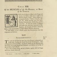 0135 - Page 35 - Chap. XIII. Of the Muscles of the Os Hyoides, or Bone of the Tongue. XLIV. Sternohyoideus
-
 0136 - Page 36 - XLV. Coracohyoideus. HLVI. Stylohyoideus. XLVII. Mylohyoideus
-
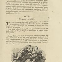 0137 - Page 37 - XLVIII. Geniohyoideus
-
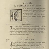 0138 - Page 38 - Chap. XIV. Of the Muscles of the Tongue. XLIX. Genioglossus. L. Ceratoglossus
-
 0139 - Page 39 - LI. Styloglossus. LII. Basioglossus
-
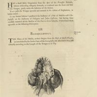
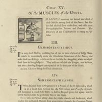 0140 - Page 40 - Chap. XV. Of the Muscles of the Uvula. LIII. Glossostaphylinus. LIV. Sphenostaphylinus
-
 0141 - Page 41 - LV. Pterygostaphylinus
-
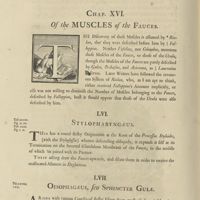 0142 - Page 42 - Chap. XVI. Of the Muscles of the Fauces. LVI. Stylopharynaeus. LVII. Oesophagaeus, seu sphincter gulae
-
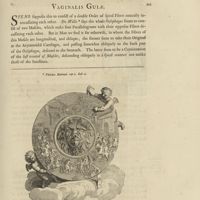 0143 - Page 43 - LVIII. Vaginalis gulae
-
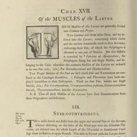 0144 - Page 44 - Chap. XVII. Of the Muscles of the Larynx. LIX. Sternothyroideus
-
 0145 - Page 45 - LX. Hyothyroideus. LXI. Cricothyroideus. LXII. Cricoarytaenoideus posticus
-
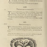 0146 - Page 46 - LXIII. Cricoarytaenoideus lateralis. LXIV. Thyroarytaenoideus. LXV. Arytaenoideus
-
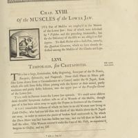 0147 - Page 47 - Chap. XVIII. Of the Muscles of the Lower Jaw. LXVI. Temporalis, seu Crotaphites
-
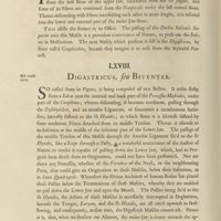 0148 - Page 48 - LXVII. Masseter. LXVIII. Digastricus, seu Biventer
-
 0149 - Page 49 - LXIX. Pterygoideus internus. LXX. Pterygoideus externus
-
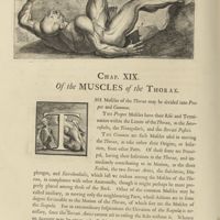 0150 - Page 50 - Chap. XIX. Of the Muscles of the Thorax
-
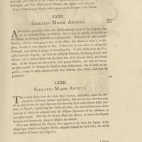 0151 - Page 51 - LXXI. Serratus minor anticus. LXXII. Serratus major anticus
-
 0152 - Page 52 - LXXIII. Subclavius. LXXIV. Scalenus primus. LXXV. Scalenus secundus
-
 0153 - Page 53 - LXXVI. Scalenus tertius. LXXVII. Triangularis. LXXVIII. Diaphragma
-
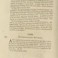 0154 - Page 54 - LXXIX. Intercostales interni
-
 0155 - Page 55 - LXXX. Cor
-
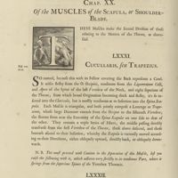 0156 - Page 56 - Chap. XX. Of the Muscles of the Scapula, or Shoulder-Blade. LXXXI. Cucullaris, seu Trapezius. LXXXII. Rhomboides
-
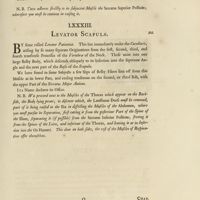 0157 - Page 57 - LXXXIII. Levator Scapulae
-
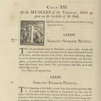 0158 - Page 58 - Chap. XXI. Of the Muscles of the Thorax, which appear on the backside of the Body. LXXXIV. Serratus superior posticus. LXXXV. Serratus inferior posticus
-
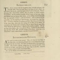 0159 - Page 59 - LXXXVI. Sacrolumbalis. LXXXVII. Intercostales externi
-
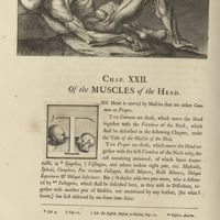 0160 - Page 60 - Chap. XXII. Of the Muscles of the Head
-
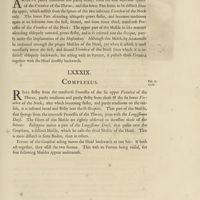 0161 - Page 61 - LXXXVIII. Splenius. LXXXIX. Complexus
-
 0162 - Page 62 - XC. Rectus major. XCI. Rectus minor. XCII. Obliquus superior. XCIII. Obliquus inferior
-
 0163 - Page 63 - XCIV. Mastoideus. XCV. Rectus internus major
-
 0164 - Page 64 - XCVI. Rectus internus minor. XCVII. Rectus lateralis
-
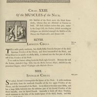 0165 - Page 65 - Chap. XXIII. Of the Muscles of the Neck. XCVIII. Longus colli. XCIX. Spinalis colli
-
 0166 - Page 66 - C. Interspinales colli. CI. Transversalis colli
-
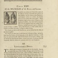 0167 - Page 67 - Chap. XXIV. Of the Muscles of the Back and Loins. CII. Longissimus dorsi
-
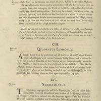 0168 - Page 68 - CIII. Quadratus lumborum. CIV. Semispinatus
-
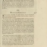 0169 - Page 69 - CV. Sacer. CVI. Musculus coccygis
-
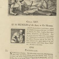 0170 - Page 70 - Chap. XXV. Of the Muscles of the Arm, or Os Humeri. CVII. Pectoralis
-
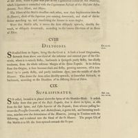 0171 - Page 71 - CVIII. Deltoides. CIX. Supraspinatus
-
 0172 - Page 72 - CX. Infraspinatus. CXI. Teres minor. CXII. Teres major. CXIII. Latissimus dorsi, sive aniscalptor
-
 0173 - Page 73 - CXIV. Coracobrachialis. CXV. Subscapularis
-
 0174 - Page 74 - Chap. XXVI. Of the Muscles of the Cubit. CXVI. Biceps
-
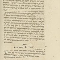 0175 - Page 75 - CXVII. Brachiaeus internus
-
 0176 - Page 76 - CXVIII. Gemellus. CXIX. Brachiaeus externus. CXX. Anconaeus
-
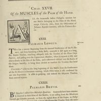 0177 - Page 77 - Chap. XXVII. Of the Muscles of the Palm of the Hand. CXXI. Palmaris longus. CXXII. Palmaris brevis
-
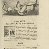 0179 - Page 79 - Chap. XXVIII. Of the Muscles of the four Fingers. CXXIII. Perforatus
-
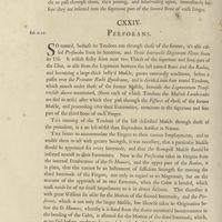 0180 - Page 80 - CXXIV. Perforans
-
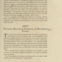 0181 - Page 81 - CXXV. Lumbricales. CXXVI. Extensor Digitorum communis, seu Digitorum tensor
-
 0182 - Page 82 - CXXVII. Interossei manus. CXXVIII. Extensor indicis, seu indicator. CXXIX. Abductor indicis
-
 0183 - Page 83 - CXXX. Extensor minimi digiti. CXXXI. Abductor minimi digiti
-
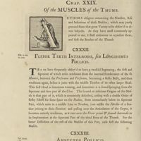 0184 - Page 84 - Chap. XXIX. Of the Muscles of the Thumb. CXXXII. Flexor tertii internodii, seu longissimus pollicis. CXXXIII. Abductor pollicis
-
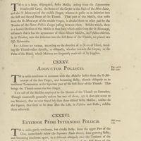 0185 - Page 85 - CXXXIV. Flexor primi & secundi ossis pollicis. CXXXV. Abductor pollicis. CXXXVI. Extensor primi internodii pollicis
-
 0186 - Page 86 - CXXXVII. Extensor secundi internodii pollicis. CXXXVIII. Extensor tertii internodii pollicis
-
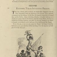
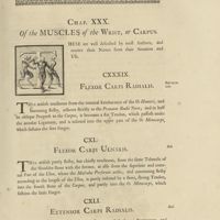 0187 - Page 87 - Chap. XXX. Of the Muscles of the Wrist, or Carpus. CXXXIX. Flexor Carpi radialis. CXL. Flexor Carpi ulnaris. CXLI. Extensor Carpi radialis
-
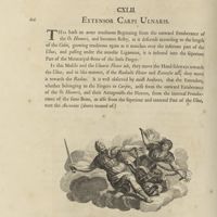 0188 - Page 88 - CXLII. Extensor Carpi ulnaris
-
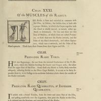 0189 - Page 89 - Chap. XXXI. Of the Muscles of the Radius. CXLIII. Pronator Radii teres. CXLIV. Pronator Radii quadratus, or inferior quadratus
-
 0190 - Page 90 - CXLV. Supinator Radii longus. CXLVI. Supinator Radii brevis
-
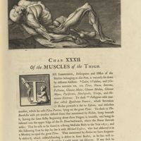 0191 - Page 91 - Chap. XXXII. Of the Muscles of the Thigh
-
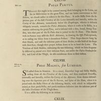 0192 - Page 92 - CXLVII. Psoas parvus. CXLVIII. Psoas magnus, seu Lumbalis. CXLIX. Iliacus internus
-
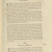 0193 - Page 93 - CL. Pectineus. CLI. Glutaeus major
-
 0194 - Page 94 - CLII. Glutaeus medius. CLIII. Glutaeus minor
-
 0195 - Page 95 - CLIV. Pyriformis, seu Iliacus externus. CLV. Marsupialis, seu Bursalis
-
 0196 - Page 96 - CLVI. Quadratus Femoris. CLVII. Obsturator externus. CLVIII. Triceps
-
 0198 - Page 98 - Chap. XXXIII. Of the Muscles of the Leg. CLIX. Membranosus
-
 0199 - Page 99 - CLX. Sartorius. CLXI. Gracilis
-
 0200 - Page 100 - CLXII. Biceps. CLXIII. Seminervosus, seu Semitendinosus. CLXIV. Semimembranosus
-
 0201 - Page 101 - CLXV. Popliteus. CLXVI. Rectus Femoris. CLXVII. Vastus externus. CLXVIII. Vastus internus
-
 0202 - Page 102 - CLXIX. Crureus, seu Femoreus
-
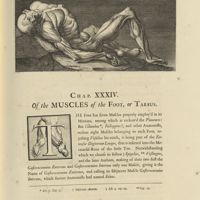 0203 - Page 103 - Chap. XXXIV. Of the Muscles of the Foot, or Tarsus
-
 0204 - Page 104 - CLXX. Tibialis Anticus. CLXXI. Gasterocnemius externus, seu Gemellus
-
 0205 - Page 105 - CLXXII. Plantaris. CLXXIII. Soleus, seu Gasterocnemius internus
-
 0206 - Page 106 - CLXXIV. Peronaeus primus. CLXXV. Peronaeus secundus
-
 0207 - Page 107 - CLXXVI. Tibialis posticus
-
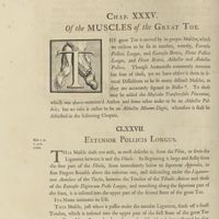 0208 - Page 108 - Chap. XXXV. Of the Muscles of the Great Toe. CLXXVII. Extensor Pollicis longus
-
 0209 - Page 109 - CLXXVIII. Extensor Pollicis brevis. CLXXIX. Flexor Pollicis longus. CLXXX. Flexor Pollicis brevis. CLXXXI. Adductor Pollicis
-
 0210 - Page 110 - CLXXXII. Abductor Pollicis
-
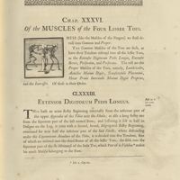 0211 - Page 111 - Chap. XXXVI. Of the Muscles of the four Lesser Toes. CLXXXIII. Extensor Digitorum Pedis longus
-
 0212 - Page 112 - CLXXXIV. Extensor Digitorum brevis. CLXXXV. Perforatus. CLXXXVI. Perforans
-
 0213 - Page 113 - CLXXXVII. Lumbricales. CLXXXVIII. Abductor minimi Digiti. CLXXXIX. Transversalis Pedis. CXC. Flexor primi internodii minimi Digiti proprius
-
 0214 - Page 114 - CXCI. Interossei Pedis
-
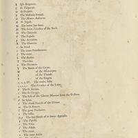
 0215 - Page 115 - Explanation of the Tables
-
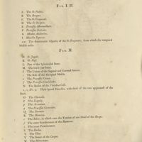
 0216 - Page 116 - Tab. I. Represents the Muscles appearing on the fore Side of a human Body, after the external Teguments are removed / Tab. II. The same body, with some of the external Muscles raised, and others taken off / Tab. III. Shews the same Body farther denuded
-
 0225 - Page 117 - Tab. IV. Shews the fore Part of the Skeleton of a Man
-
 0229 - Page 119 - Tab. V. Represents the Body in a Side View. Fig. I. and Fig. II
-
 0233 - Page 121 - Tab. VI. Represents the Side View of the Body partly denuded, and the Skeleton. Fig. I. and Fig. II
-
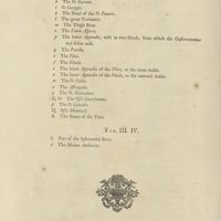 0234 - Page 122 - Fig. III and Fig. IV
-
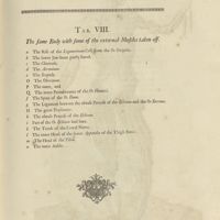 0239 - Page 123 - Tab. VII. Shews the back View of the Body a Man / Tab. VIII. The same Body with some of the external Muscles taken off
-
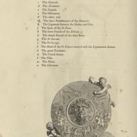 0240 - Page 124 - Tab. IX. Represents the same Body, with more of the Muscles removed
-
 0245 - Page 125 - Tab. X. The back View the Skeleton of a Man
-
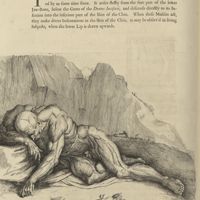
 0246 - Page 126 - Tab. XI. XII. XIII. These Figures are chiefly design'd for the Use of Painters and Statuaires. The Outlines are copied from some of the greatest Masters, and the Muscles are laid in from the Life
-
 0257 - Page 127 - Tab. XIV. Shews the internal Surface of the Musculus Obliquus Descendens, when dissected and display'd
-
 0258 - Page 128 - Tab. XV. The Musculus Obliquus Ascendens, with the Rectus and part of the Tendon of the Descendens, together with the Testis of the Left Side dissected and display'd...
-
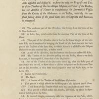
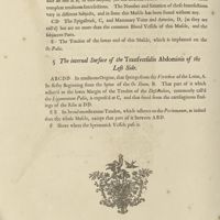 0262 - Page 130 - Tab. XVI. 4. The internal Surface of the Rectus Abdominis of the Left Side. 5. The internal Surface of the Transversalis Abdominis of the Left Side
-
 0267 - Page 131 - Tab. XVII
-
 0268 - Page 132 - Tab. XVIII
-
 0273 - Page 133 - Tab. XIX
-
 0274 - Page 134 - Tab. XX
-
 0279 - Page 135 - Tab. XXI
-
 0280 - Page 136 - Tab. XXII
-
 0285 - Page 137 - Tab. XXIII
-
 0286 - Page 138 - Tab. XXIV
-
 0291 - Page 139 - Tab. XXV. The Muscles of the Eye-lid and Eye, Ala Nasi & Auricula, as big as the Life. Fig. I. The Muscles of the Palpebra, and right Eye in self, dissected and display'd. Fig. II. The back part of the Muscles of the Left Eye dissected and display'd. Fig. III. IV. The Muscles of the Eye dissected, and so dispos'd, as to shew the natural Position ef each Muscle in the Left Eye
-
 0292 - Page 140 - Fig. V. VI. Fig. VII. The Left Ear, with its Muscles, as I have most commody found them, they varying in their Figure in different Subjects
-
 0295 - Page 141 - Tab. XXVI. Fig. I. Part of the Right Os Temporale, &c. containingh the Organs of Hearing of the Right Side as big as the Life. Fig. II. The internal Surface of the same Bones, with the Organs of Hearing, represented in the preceding Figure
-
 0296 - Page 142 - Fig. III. Part of preceding Figure. Fig. IV. The four little Bones of the roght Ear taken out with their Muscles
-
 0297 - Page 143 - Fig. V. VI. VII. The Malleus, Incus, and Stapes, as big as the Life, and magnisy'd. Fig. VIII. The same Organs of the Left Ear magnify'd in the same Position, as in Fig. III
-
 0298 - Page 144 - Tab. XXVII. Fig. I. The upper Surface of the Os Hyoides, when freed from its Muscles. Fig. II. The internal Surface of the same Os Hyoides. Fig. III. IV. The internal and external Surface of the Os Hyoides from another Body. The two other Figures represent the Os Hyoides taken out with its Muscles
-
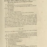 0303 - Page 145 - Tab. XXVIII. Fig. I. The Tongue, with its Muscles cut from their Originations, and left at their Insertions into the Body of the Tongue. Fig. II. The back part of the Muscles of the Uvula, with the upper part of those of the Fauces, when dissected
-
 0304 - Page 146 - Fig. III. The back part of the Muscles of the Fauces, connected to the Os Hyoides and Larynx
-
 0305 - Page 147 - Fig. IV. The Muscles of the Uvula, clear'd and left in their natural Situation
-
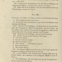
 0306 - Page 148 - Tab. XXIX. Fig. I. II. The fore Parts of the Muscles of the Uvula and Fauces dissected and cleared of their Glandulous Membranes. Fig. III. The Gula distended with Wind, to shew. Fig. IV. The Musculus Vaginalis Gulae, ending on the Left Orifice of the Stomach
-
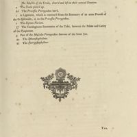
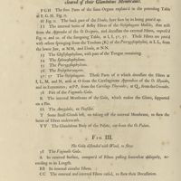
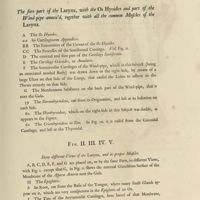 0311 - Page 149 - Tab. XXX. Fig. I. The fore part of the Larynx, with the Os Hyoides and part of the Wind pipe annex'd, together all the common Muscles of the Larynx. Fig. II. III. IV. V. Shew different Views of the Larynx, and its proper Muscles
-
 0315 - Page 151 - Tab. XXXI. The Muscles of the lower Jaw, as they appear, when freed from the Cranium
-
 0316 - Page 152 - Tab. XXXII
-
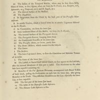
 0321 - Page 153 - Tab. XXXIII. Fig. I. The Inside of the Sternum, with the Cartilaginous Endings of the Ribs, freed from their Bony Parts. Fig. II. A Sketch of the internal Surface of two or three of the lower Ribs, near their Articulations with the Vertebrae of the Back, as big as the Life; where a Disposition of the internal Intercostal Muscles, not commonly taken notice of, is represented
-
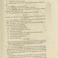
 0322 - Page 154 - Tab. XXXIV. The upper Surface of the Diaphragm, next the Thorax
-
 0327 - Page 155 - Tab. XXXV. The lower Surface of the Diaphragm, next the Abdomen
-
 0328 - Page 156 - Tab. XXXVI. Divers Views of the external, middle, and internal Compages of the Fibres of the Heart. Fig. I. The fore part of the Heart, next the Sternum. Fig. II. The back part of the Heart, next the Vertebrae
-
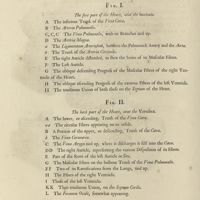
 0331 - Page 157 - Fig. III. The Series of Fibres, under those represented in Fig. I Fig. IV. The Fibres, as they appear, under those express'd in Fig. III. Fig. V. The Double Spiral Order of the Fibres, at the Cone of the Heart, which may partly be seen in Fig. III. Fig. VI. A View of the internal Surface of the Cone of the Heart, after a transverse Section
-
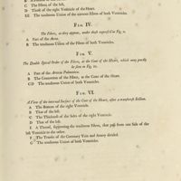
 0332 - Page 158 - Tab. XXXVII. Fig. I. The right Auricle and Ventricle of the Heart, open'd from the Vena Cava Fig. II
-
 0335 - Page 159 - Fig. III
-
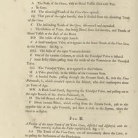
 0336 - Page 160 - Tab. XXXVIII. Fig. I. The same Heart, represented at Fig. I. in the preceding Table, with the right Ventricle open'd from the Arteria Pulmonalis, both being here pinn'd out and display'd, as big as the Life. Fig. II. The Mitral Valve of the left Ventricle, dissected and display'd
-
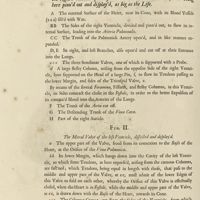
 0339 - Page 161 - Fig. III. The internal Surface of the three Semilunar Valves of the Pulmonary Artery, next the Cavity of the right Ventricle of the Heart. Fig. IV. The external Surface of the same Valves, next the Trunk of the Pulmonick Artery
-
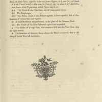
 0340 - Page 162 - Tab. XXXIX. Fig. I. The Left Ventricle of the Heart open'd, with the Trunk of the Vena Pulmonalis, and the Left Auricle. Fig. II. The external Paries of the Left Ventricle, cut off from the Basis of the Heart, at the Root of the Arteria Magna, to the Cone, according to the length of the Heart
-
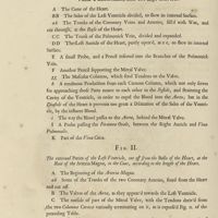
 0343 - Page 163 - Fig. III. The Mitral Valve taken out entire, with the Columnae Carneae and their annex'd Tendons, &c.
-
 0344 - Page 164 - Tab. XL. Fig. I. The Left Ventricle of the Heart opened, together with the Vena Pulmonalis and Trunk of the Aorta, to shew in one View the Position of the Valves, that hinder the Return of the Blood by the same way it came into the Ventricle, as well as those Valves, that prevent the return of the Blood, after it is expell'd from thence by the Systole of the Heart. Fig. II. Is drawn from the same Heart express'd Fig. I. in the preceding Table, after dividing the Vena Pulmonalis, through the middle, (as 'tis there represented) together with the Arteria Aorta
-
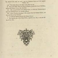
 0347 - Page 165 - Fig. III. Part of the Aorta cut off next the Basis of the Heart, to shew its Valves, as they appear next the Ventricle, when the Heart is in Diastole, and the Blood hinder'd by them in its return from the Aorta to the Left Ventricle, after its Expulsion from thence
-
 0348 - Page 166 - Fig. IV. A Portion of the Aorta, with the Parts annex'd, in the same Position, as was represented in the preceding Figure, taken from a morbid Body, in which the use of the Semilunar Valves was entirely defeated by a large Petrefaction, that grew upon them. Fig. V. VI. Shew the same Parts of the Aorta, with its Valves viewed from the great Artery towards the Heart
-
 0351 - Page 167 - Tab. XLI
-
 0352 - Page 168 - Tab. XLII
-
 0357 - Page 169 - Tab. XLIII
-
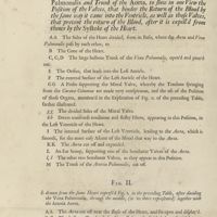
 0358 - Page 170 - Tab. XLIV. Fig. I. The back Parts of the Vertebrae of the Neck, as they appear'd in a late Subject, with the Muscles and Parts annex'd, as big as the Life
-
 0362 - Page 172 - Tab. XLV. Fig. I. II. The exteriour and interiour Surfaces of the Longissimus Dorsi, Sacrolumbalis, Sacer, Semi-Spinatus, and Transversalis Colli, dissected from an emaciated Body
-
 0368 - Page 174 - Tab. XLVI / Tab. XLVII
-
 0373 - Page 175 - Tab. XLVIII
-
 0374 - Page 176 - Tab. XLIX
-
 0379 - Page 177 - Tab. L
-
 0380 - Page 178 - Tab. LI
-
 0385 - Page 179 - Tab. LII
-
 0386 - Page 180 - Tab. LIII
-
 0390 - Page 182 - Tab. LIV
-
 0397 - Page 183 - Tab. LV. LVI.
-
 0398 - Page 184 - Tab. LVII
-
 0403 - Page 185 - Tab. LVIII
-
 0404 - Page 186 - Tab. LIX
-
 0409 - Page 187 - Tab. LX
-
 0410 - Page 188 - Tab. LXI
-
 0415 - Page 189 - Tab. LXII
-
 0416 - Page 190 - Tab. LXIII. The Muscles of the Toes
-
 0421 - Page 191 - Tab. LXIV. The Muscles of the Toes, as big as the Life
-
 0422 - Page 192 - Tab. LXV
-
 0426 - Page 194 - Tab. LXVI
-
![0432 - Page sans numérotation - [Page de garde]](https://numerabilis.u-paris.fr/iiif/2/bibnum:01892:0432/square/200,/0/default.jpg) 0432 - Page sans numérotation - [Page de garde]
0432 - Page sans numérotation - [Page de garde]
-
![0433 - Page sans numérotation - [Contreplat]](https://numerabilis.u-paris.fr/iiif/2/bibnum:01892:0433/square/200,/0/default.jpg) 0433 - Page sans numérotation - [Contreplat]
0433 - Page sans numérotation - [Contreplat]
-
![0434 - Page sans numérotation - [Plat]](https://numerabilis.u-paris.fr/iiif/2/bibnum:01892:0434/square/200,/0/default.jpg) 0434 - Page sans numérotation - [Plat]
0434 - Page sans numérotation - [Plat]
- Sur l'auteur
- Cowper, William (1666 - 1709)
- Identifiant SUDOC
- Notice dans le Sudoc